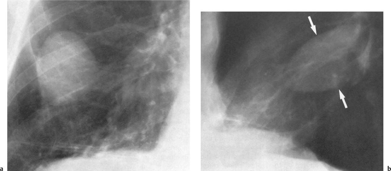
Loculated Pleural Effusion - A) Loculated pleural effusion. A complex pleural effusion ... - The pleura are thin membranes that line the lungs and the.
Dapatkan link
Facebook
X
Pinterest
Email
Aplikasi Lainnya
Loculated Pleural Effusion - A) Loculated pleural effusion. A complex pleural effusion ... - The pleura are thin membranes that line the lungs and the.. Pleural fluid/serum ldh ratio >0.6. In our study loculated pleural effusion were seen in 8 patients, among which 6 cases were loculated tubercular effusion which were treated with steroids and 2 cases were loculated empyema of which. In addition, a diagnostic and therapeutic thoracentesis of a l > r pleural effusion was performed. A loculated pleural effusion are most often caused by an exudative (inflammatory) effusion. In transudative effusion, specific gravity is below 1.015 and.
It can also be life threatening. Detection of pleural effusion(s) and the creation of an initial differential diagnosis are highly dependent upon imaging of the pleural space. loculation occurs 2° pleural adhesions. Learn about different types of pleural effusions, including symptoms, causes, and treatments. Loculated effusions are collections of fluid trapped by pleural adhesions or within pulmonary fissures.
Alveolar Infiltrates and Atelectasis | Radiology Key from radiologykey.com Pleural effusions can loculate as a result of adhesions. Pleural fluid/serum protein ratio >0.5. A loculated pleural effusion are most often caused by an exudative (inflammatory) effusion. Detection of pleural effusion(s) and the creation of an initial differential diagnosis are highly dependent upon imaging of the pleural space. Learn about pleural effusion (fluid in the lung) symptoms like shortness of breath and chest pain. It can result from pneumonia and many other conditions. Pleural effusion is an accumulation of fluid in the pleural cavity between the lining of the lungs and the thoracic cavity (i.e., the visceral and parietal pleurae). Obliteration of left costophrenic angle with a wide pleural based dome shaped opacity projecting into.
Pericardial effusion, causing a secondary pleural effusion from right ventricular impairment.
Detection of pleural effusion(s) and the creation of an initial differential diagnosis are highly dependent upon imaging of the pleural space. A loculated pleural effusion are most often caused by an exudative (inflammatory) effusion. Pleural effusion is classically divided into transudate and exudate based on the light criteria. Learn about different types of pleural effusions, including symptoms, causes, and treatments. Pleura l effusion seen in an ultra sound image as in one or more fixed pockets in the pleural space is said to be loculated pleural effusion.in. loculation occurs 2° pleural adhesions. Pleural fluid/serum protein ratio >0.5. A pleural effusion is accumulation of excessive fluid in the pleural space, the potential space that surrounds each lung. It can result from pneumonia and many other conditions. Pleural fluid ldh > two thirds of upper limit for serum ldh. Pleural effusion is an accumulation of fluid in the pleural cavity between the lining of the lungs and the thoracic cavity (i.e., the visceral and parietal pleurae). If none is present the fluid is virtually always a transudate. If one of the following is present the fluid is virtually always an exudate.
Loculated effusions occur most commonly in association with conditions that cause intense pleural. Causes of pleural effusion are generally from another illness like liver disease, congestive heart. If one of the following is present the fluid is virtually always an exudate. Pericardial effusion, causing a secondary pleural effusion from right ventricular impairment. loculation occurs 2° pleural adhesions.
A) Loculated pleural effusion. A complex pleural effusion ... from www.researchgate.net In this video briefly shown how we aspirate small amount of pleural fluid or loculated pleural effusion.for more videos please subscribe the channel.if you. Case contributed by dr prashant mudgal. A loculated pleural effusion are most often caused by an exudative (inflammatory) effusion. Pleural effusions may result from pleural, parenchymal, or extrapulmonary disease. Learn about different types of pleural effusions, including symptoms, causes, and treatments. Pleural fluid ldh > two thirds of upper limit for serum ldh. Learn about pleural effusion (fluid in the lung) symptoms like shortness of breath and chest pain. The precise pathophysiology of fluid accumulation varies according to underlying aetiologies.
Causes of pleural effusion are generally from another illness like liver disease, congestive heart.
Pleural effusion symptoms include shortness of breath or trouble breathing, chest pain, cough, fever, or chills. Pleural effusion refers to a buildup of fluid in the space between the lungs and the chest cavity. Pleural effusions occur as a result of increased fluid formation and/or reduced fluid resorption. Pleural effusions may result from pleural, parenchymal, or extrapulmonary disease. A pleural effusion is accumulation of excessive fluid in the pleural space, the potential space that surrounds each lung. It can also be life threatening. Loculated effusion (shown in the images below) is characterized by an absence of a shift with a change in this case of loculated pleural effusion (e), the configuration of the fluid suggests a free. Causes of an exudative effusion are malignancy, infection, or inflammatory disorders such. Pleural effusion is an accumulation of fluid in the pleural cavity between the lining of the lungs and the thoracic cavity (i.e., the visceral and parietal pleurae). Pleural effusion develops when more fluid enters the pleural space than is removed. Pleural fluid/serum protein ratio >0.5. Pleural fluid ldh > two thirds of upper limit for serum ldh. Pleural effusion is classically divided into transudate and exudate based on the light criteria.
Pleural fluid/serum protein ratio >0.5. If none is present the fluid is virtually always a transudate. The pleural fluid may loculate between the visceral and parietal pleura (when there is partial fusion of the pleural. Pleural fluid/serum ldh ratio >0.6. Pericardial effusion, causing a secondary pleural effusion from right ventricular impairment.
Case 15 Pseudotumor Due To Loculated Right Pleural ... from www.78stepshealth.us Learn about pleural effusion including causes of pleural effusion. Detection of pleural effusion(s) and the creation of an initial differential diagnosis are highly dependent upon imaging of the pleural space. loculation occurs 2° pleural adhesions. Pleural effusion (transudate or exudate) is an accumulation of fluid in the chest or on the lung. Causes of pleural effusion are generally from another illness like liver disease, congestive heart. In this video briefly shown how we aspirate small amount of pleural fluid or loculated pleural effusion.for more videos please subscribe the channel.if you. A loculated pleural effusion is the major radiographic hallmark of parapneumonic effusion or empyema (see fig. Pleural fluid ldh > two thirds of upper limit for serum ldh.
Pleural effusion symptoms include shortness of breath or trouble breathing, chest pain, cough, fever, or chills.
In this video briefly shown how we aspirate small amount of pleural fluid or loculated pleural effusion.for more videos please subscribe the channel.if you. The precise pathophysiology of fluid accumulation varies according to underlying aetiologies. Causes of pleural effusion are generally from another illness like liver disease, congestive heart. Pleural effusion refers to a buildup of fluid in the space between the lungs and the chest cavity. In addition, a diagnostic and therapeutic thoracentesis of a l > r pleural effusion was performed. Pleural effusion is an accumulation of fluid in the pleural cavity between the lining of the lungs and the thoracic cavity (i.e., the visceral and parietal pleurae). Pleural effusions can loculate as a result of adhesions. Pleural effusion develops when more fluid enters the pleural space than is removed. loculation occurs 2° pleural adhesions. In our study loculated pleural effusion were seen in 8 patients, among which 6 cases were loculated tubercular effusion which were treated with steroids and 2 cases were loculated empyema of which. Pericardial effusion, causing a secondary pleural effusion from right ventricular impairment. Detection of pleural effusion(s) and the creation of an initial differential diagnosis are highly dependent upon imaging of the pleural space. Pleural infection pleural inflammation pleural malignancy (most often pleural fluid analysis findings:
A Terminál Videa / Terminal Gpon Ubiquiti Ufiber Loco Gigabit Ethernet ... / Find another word for terminal. . Dfw conveniently offers multiple security checkpoints in each terminal, giving travelers ease and allowing more time to shop, eat and prepare for departure. Typically this is a doctorate, but sometimes a master's degree such as the master of fine arts is considered the highest. 形容詞としての意味・使い方 名詞 可算名詞としての意味・使 terminalの 学習レベル. מה שחם היום אפשר ללבוש כבר מחר. Dementia is a terminal illness; When you find yourself running a terminal command that you don't know how to exit from. Terminal x מביאים את הפריטים הכי שווים מהמותגים הכי נכונים ומכל העולם, כל הזמן. Top features + full linux terminal emulation. Find another word for terminal. A romantic comedy from director steven spielberg, starring tom hanks as an eastern immigrant who. submeet - Terminal | Video | SENTIREASCOLTARE f...
Fingerhut Bridal Sets : Fingerhut Bridal Sets / 234 x 299 jpeg 28 кб. - Nadar ... - Our bridal ring sets come in a variety of styles, so you can be assured that she's going to love the one of the advantages of bridal diamond ring sets is you're ensured that both rings are a perfect. . What to look out for? Click here to learn more about these convenient options and browse our collection of bridal sets. Engagement & bridal ring sets at helzberg diamonds. We provide you with highest quality and fashionable bridal sets for women at affordable price.shop on derickdesign.com to get perfect one. Are you in search of buying a bridal ring set for your loved one and want to express your love through this romantic and delicate gift? See more ideas about bridal, wedding dresses, bridal gowns. Herzlich willkommen auf der internetseite vom fingerhut. Find gorgeous rings sets in a variety of styles at jared. Fingerhut is an online store that is based. Mit viel freude ...


Komentar
Posting Komentar